Unit 1.1: Investigating an Outbreak
|
In the first section of the Unit, our class was given a fake family with many ailments, from infections to diseases, and we were given the task of finding out what infection Sue, the daughter in college, got while in college. Each student is the chief investigator in the case, and we had to use the given patient symptoms and medical histories to investigate the outbreak.
We had to find possible diseases responsible for the outbreak, and we narrowed down our search with multiple diagnostic techniques. My classmates and I used DNA sequence analysis to identify the illness. In the first unit, we will not only perform an ELISA experiment, but we will also take the given information and put it into a chart to help determine who and where the disease came from. |
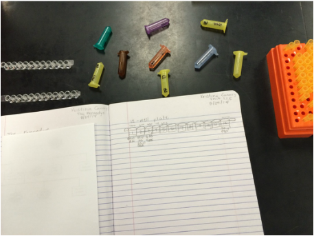
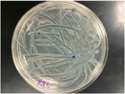
Ampicillin, streptomycin, ecoli I and II control, and strep and amp mix plates, as well as other tools used in experiment for unit 1.2.3
|
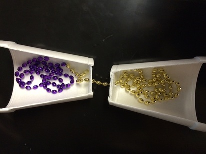
Representation of how a bacterial cell transfers DNA from one another using either conjugation, transformation, and transduction. The picture above is of transduction. The beads represent the seperate (now combining) DNA, and the cups represent the separate bacterium.
|
Unit 1.2: Antibiotic Therapy
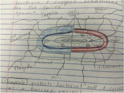
In this activity of the unit, my peers and I reviewed the structure of bacterial cells, and learned about the various types of antibiotics and their modes of action against the bacteria it affects. We learned about the effectiveness of antibiotics against certain strains of bacteria, and how they're dependent on the mechanism of action and structure of the bacteria.
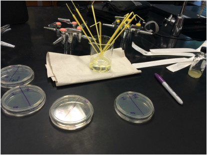
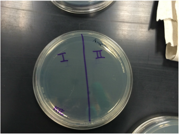
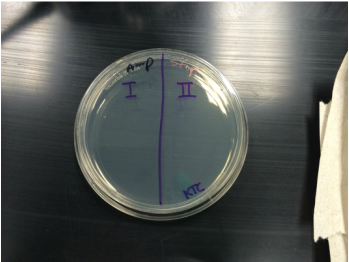
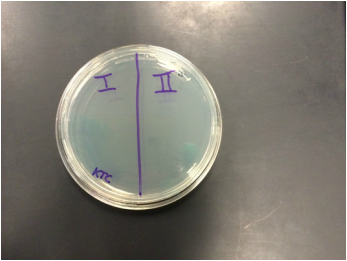
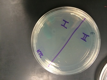
We used three agar plates, which either held streptomycin bacteria, ampicillin bacteria, and then both bacteria on each individual plate. There were two given strains of E coli (I & II), which were kept separated on all four plates before the incubation period. Each E coli strain had certain traits within their DNA which helped them in survival of certain antibiotics. The picture above is a gram positive and gram negative bacterial cell.
We used three agar plates, which either held streptomycin bacteria, ampicillin bacteria, and then both bacteria on each individual plate. There were two given strains of E coli (I & II), which were kept separated on all four plates before the incubation period. Each E coli strain had certain traits within their DNA which helped them in survival of certain antibiotics. The picture above is a gram positive and gram negative bacterial cell.
Ampicillin plate before incubation
Strep and Amp plate before incubation
|
Streptomycin plate before incubation
Control plate with only Ecoli cultures I & II before incubation
|
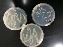
After the plates were incubated, all students and their partners had to observe the bacterial growth that took place with each E coli strain on each agar plate. We then recorded the growth in a table which stated that E coli I survives on the strep plate, but doesn't in the amp and the mixed plate. While E coli II could survive on the amp plate but does not on the strep and the mixed plate.
My partner and I then took a colony from the Ecoli I colony on the strep plate, and then a colony from the Ecoli II strain on the amp plate. We then streaked the colonies along the surface of the agar plate.
Then, we took another three plates of ampicillin, streptomycin, and antibiotic mix plate to test the effectiveness of the mix of E coli I & II antibiotic resistant bacteria. This was to see that DNA was properly shared among the colonies to create a "Super Bug" that is resistant to both ampicillin and streptomycin.
After incubation of E coli I strep resistant & E coli II amp resistant mix plate
|
After incubation of amp and strep resistant bacterial colony on strep, amp, and mix plates
|
The students then made the 3D model (as shown in Unit 1.1), which could either represent transduction, conjugation, or transformation. Our model shows transduction, which is when DNA is transferred by another virus.
Unit 1.3: Can You Hear Me Now?
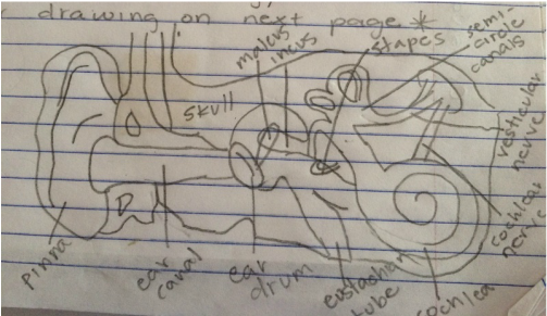
Inner ear drawing
Parts of the Ear:
1. Pinna- The function of the pinna is to collect sound as a canal, amplify it, and then direct it to the auditory canal.
2. Auditory canal- The function of the auditory canal is for helping the auditory process by funneling sound toward the eardrum and protecting it from injury.
3. Eustachian tube- Its function is to connect the middle ear cavity and the nasopharynx, while also clearing mucus and aerating the middle ear system.
4. Ossicles (malleus, incus, & stapes)- Its functions are to transmit sounds from the air to the fluid-filled labyrinth (cochlea).
5. Tympanic membrane (eardrum)- Its function is to seperate the external and middle ear and transmit sound from air to the ossicles inside the middle ear.
6. Cochlea- The cochlea transforms the vibrations of the cochlear liquids and associated structures into neural signal.
7. Sensory hair cells- Its function is to amplify sound waves and transduce auditory information to the brain stem.
8. Cochlear nerve- The cochlear nerve carries sound waves from the cochlea of the inner ear to the brain.
9. Oval window- The oval window is a membrane-covered opening which leads from the middle ear to the inner ear where sound vibrations are transmitted.
10. Vestibule- The vestibule connects the cochlea and semicircular canals and contains two other balance and equilibrium related structures.
11. Vestibular nerve- Its function is to carry sensory information related to body equilibrium as part of the auditory nerve.
Unit 1.3: Continued...
In this activity, Sue has recovered from her bacterial meningitis, but was recommended to see a physician about her hearing because of the high rate of hearing loss in meningitis patients. We investigated the physics of sound and learned about how hearing works so that we may effectively diagnose a patient with certain types of hearing loss.
We created a model of the ear to better understand the anatomy of how certain hearing losses affect certain parts. My partner and I also performed a Rhine test and audio grams to help understand how to correctly diagnose patients, as well as test our own hearing as an example.
The Rhine test is used with the tuning fork, which was hit on the sole of your shoe and your partner was to tell when the vibration on his bone behind his ear ceased and then when the sound stopped afterwards. Next, we were to plug in a pair of headphones and conduct a hearing test, which utilized logger pro, and sounded a different sound every time you changed the hertz. You were to start at the highest volume of sound, and then gradually subtract the sound until you can't hear it anymore. This was measured on a graph to test your hearing at different hertz in both ears.
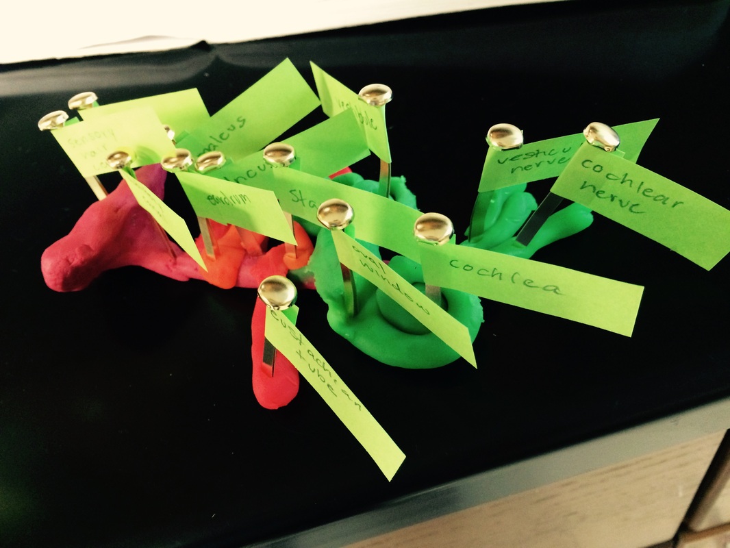
Labeled inner ear model
|
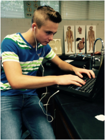
Kaleb Decker with headphones as he conducts his hearing test
|

Kaleb Decker with the tuning fork which was used to conduct vibrations to test bone and air conduction in each ear
|
Unit 1.4: Vaccinations
A vaccine is a preparation of killed or weakened microorganisms that is administered to produce or artificially increase immunity to a particular disease. There several different types of vaccines including: killed, weakened, toxoid, which uses toxins from the antigen, subunit, whish uses recombinant DNA technology, naked DNA, and similar pathogen vaccines.
One shot against influenza doesn't necessarily protect a person from year to year because influenza mutates very rapidly into many different strains, so one flu shot may protect you from the flu that year, but by next year, its mutated into another strain so your shot from last year wouldn't protect you.
One shot against influenza doesn't necessarily protect a person from year to year because influenza mutates very rapidly into many different strains, so one flu shot may protect you from the flu that year, but by next year, its mutated into another strain so your shot from last year wouldn't protect you.

|
This is a diagram of plasmid in the form of a ring, with plasmid A & B connected by viral DNA. It represents how a virus's DNA would incorporate itself into the original plasmids DNA. |
1.4.3: Life of an Epidemiologist
An Epidemiologist does field work to determine what causes disease or injury, what the risks are, who is at risk, and how to prevent further incidences. They also understand demographic and social trends that result from disease and injury.
An epidemiologists analysis helps to establish if some kind of behavior of the disease correlates to the condition, and if the correlation is a risk factor for that disease. Examples would be if smoking can have a correlation to having lung cancer, or if obesity can cause heart disease.
An epidemiologists analysis helps to establish if some kind of behavior of the disease correlates to the condition, and if the correlation is a risk factor for that disease. Examples would be if smoking can have a correlation to having lung cancer, or if obesity can cause heart disease.
